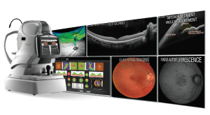
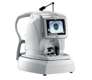
NIDEK OCT RS-330 RETINA SCAN DUO

Easy operation with 3-D auto tracking, auto shot, and user friendly interface:
The acclaimed 3-D auto tracking and auto shot functions allow easy imaging of the fundus and all its features. Each standard and professional mode has a different image capture interface which can be selected based on clinic preference.
High definition images:
For OCT imaging, up to 50 images can be averaged and the OCT sensitivity is selectable among ultra fine, fine, and regular sensitivities based on ocular pathology. The Retina Scan Duo™ has a built-in 12-megapixel CCD camera, producing high quality fundus images.
Wide area scan / Wide area normative database:
A 12 x 9 mm wide area image centered on the macula can be captured with the Retina Scan Duo™. The 9 x 9 mm normative database provides a color-coded map indicating distribution range of the patient’s macular thickness in a population of normal eyes.
Features:
- Easy operation with 3-D auto tracking, auto shot, and user friendly interface
- High definition images
- Wide area scan (12 x 9 mm) / Wide area normative database (9 x 9 mm)
- Multiple OCT scan patterns
- Value added features
- Various reports
NIDEK OCT RS-3000 ADVANCE 2

Real time compensation for eye movement with SLO-based eye tracer results in more accurate scans, ensuring higher image quality and maximum reproducibility. Selection of the appropriate OCT sensitivity allows acquisition of B-scan images through media opacities.
Features:
- Providing a comprehensive solution for retina and glaucoma analysis
- Accurate image capture with a SLO-based eye tracer
- Selectable OCT sensitivity that allows acquisition of B-scan images through media opacities
- Tracing HD for accurate averaging of up to 120 images
- Glaucoma analysis with wide-area normative database 9 x 9 mm
- High resolution AngioScan OCT-Angiography image
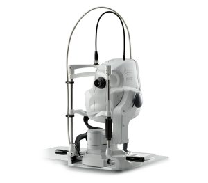
NIDEK MIRANTE SLO/OCT

The clear image of the entire 163° field of view** enables detailed evaluation of pathologies from the fovea to the extreme periphery.
* Ultra wide field imaging is available with the optional wide-field adapter.
** Measured from the center of the eye
4,096 x 4,096 pixel imaging captures every detail of the retina and choroid. Additionally, zooming in allows high magnification, clear visualization of subtle changes in pathology, and resolution of the fine details of capillaries.
New FlexTrack algorithm corrects image distortion due to unstable fixation and enhances averaging quality.
Features:
- The ultimate multimodal imaging platform
For the SLO/OCT model
– Color / FA / ICG / Blue-FAF / Green-FAF / Retro mode
– OCT / OCT-Angiography*
For the SLO model
– Color / FA* / ICG* / Blue-FAF /Green-FAF / Retro mode - Ultra wide field x ultra HD image*
- Unsurpassed color
- Dynamic/Simultaneous FA and ICG
- Unique Retro mode
- HD wide area OCT
- Fly Through function
* Optional