Overview
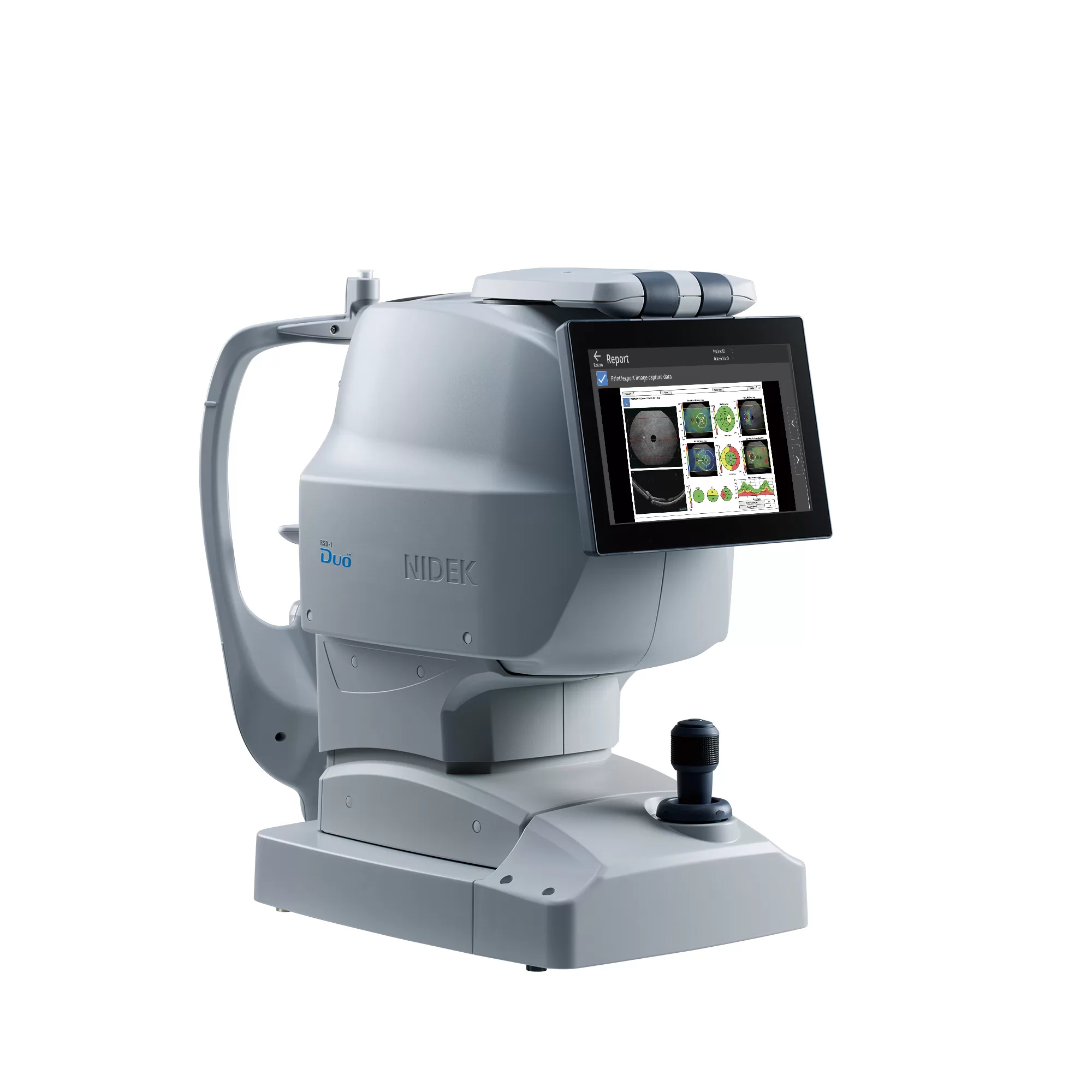
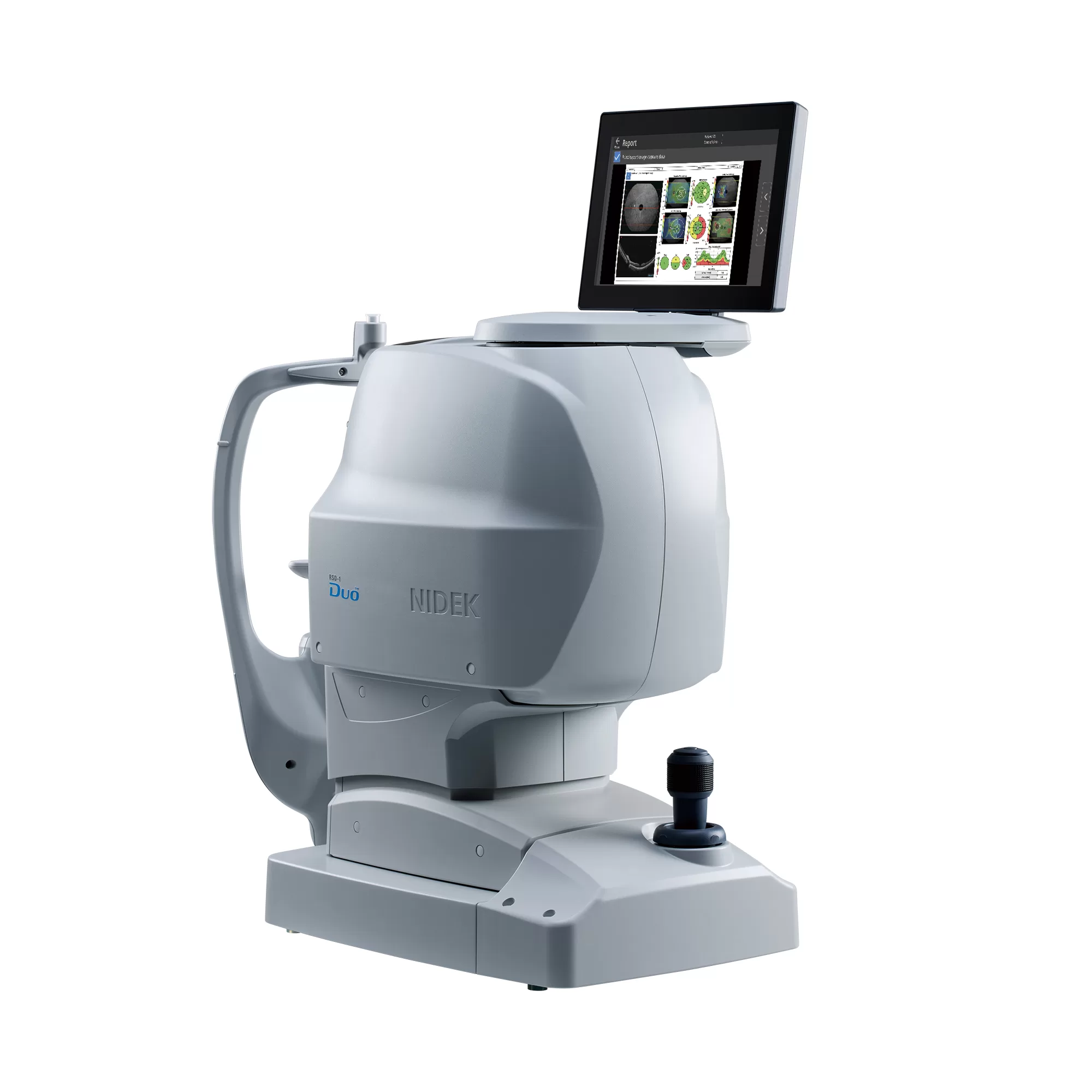
NIDEK RSD-1 Duo – Advanced OCT and Fundus Camera
The NIDEK RSD-1 Duo combines advanced Optical Coherence Tomography (OCT) and high-definition fundus photography in one compact system. Designed for efficient retinal imaging, it delivers detailed scans, automated operation, and reliable diagnostic support for modern eye care practices.
Automated and Efficient Imaging
With five automated functions, the RSD-1 Duo simplifies image acquisition and improves clinical workflow. Its intelligent automation enables quick and consistent OCT and fundus examinations.
High-Definition OCT Imaging
Featuring wide B-scans up to 16.5 mm and advanced deep-learning-based denoising, the system provides clear retinal images. The 15 × 10 mm retina map captures both the macula and optic disc for comprehensive retinal and glaucoma assessment.
Advanced Fundus Photography
The integrated slit-scan fundus camera delivers high-contrast images with a 50° field of view, supporting imaging through pupils as small as 2.0 mm.
Flexible Connectivity
The RSD-1 Duo supports DICOM connectivity and integration with NIDEK EyeLink for centralized data management and convenient examination review across multiple workstations.
With optional OCT Angiography (OCT-A) and advanced analysis capabilities, the NIDEK RSD-1 Duo provides an efficient solution for retinal imaging and glaucoma assessment.


